Resources and support for researchers
Proof-of-concept experiments using different culture systems to demonstrate their potential. To investigate new targets, proof-of-concept drug development and investigating host-pathogen interactions.
The Biomedical Imaging Unit is a joint core facility of the Faculty of Medicine and UHS providing research and diagnostic services in high quality 2D, 3D and 4D microscopy using light, electrons and X-rays for the University, NHS and external organisations. They provide training, support and expertise in imaging, correlative imaging, 3D design and printing, and image analysis.

BRAIN UK (BRain Archive Information Network UK) makes the very extensive and comprehensive NHS Neuropathology archives available to the national and international neuroscience research community. They provide access to archived biopsy and post-mortem tissue from a wide range of neurological disorders, affecting the brain and the neuromuscular system, including tumours. Digital images and clinical data are also available on request.
Clinical Samples for Patient Benefit
The Clinical Samples for Patient Benefit initiative advises on, and enables release of, consented excess pathological tissue for research purposes. For more information, please contact researchhistology@uhs.nhs.uk or Ruth Challis ruth.challis@uhs.nhs.uk
Flow Cytometry is a powerful technique which can rapidly analyse and sort millions of cells. This technique allows scientists to determine different cell populations within a sample. Analysis of the cell characteristics shows how the number and types of cells change in different diseases or treatment conditions.
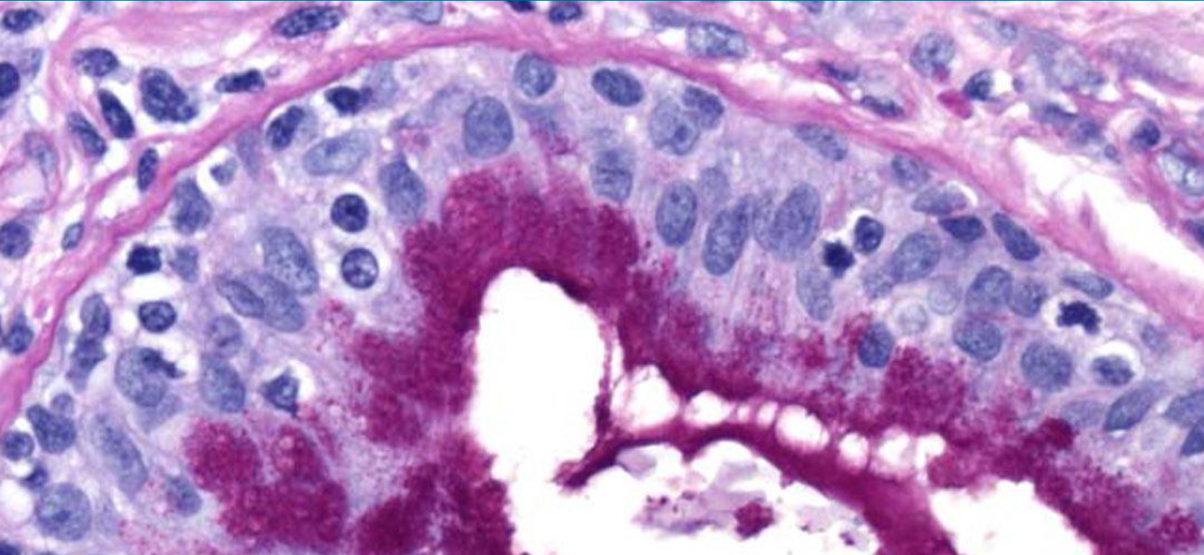
We process cells and tissue samples into a range of mediums including frozen, paraffin, and resin. Allowing sectioning and staining techniques. We can offer advice and training on a variety of techniques, and have extensive experience in histopathology for clinical trials.
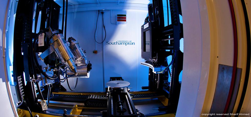
The µ-VIS X-Ray Imaging Centre is University of Southampton’s dedicated centre for microfocus Computed Tomography (µCT) and founding partner of the National Research Facility for lab-based X-ray CT (NXCT). The centre combines state-of-the-art equipment and 25 years of experience, plus the expertise of over 40 academic staff from across the university. µ-VIS constitutes a strategic multimillion pound investment in high resolution X-ray tomographic imaging, offering a unique user experience for advanced 3D imaging.
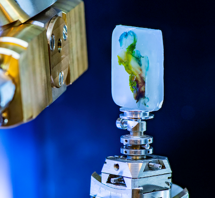
The 3D X-ray Histology facility is a pioneering platform in the field of histology and biomedical imaging. It operates within μ-VIS and the BIU, situated at the Southampton General Hospital. Specialising in non-destructive 3D imaging workflows using X-ray microfocus Computed Tomography (μCT), it is uniquely equipped for whole-block imaging of tissue specimens. The facility seamlessly integrates correlative imaging techniques, including classical histology, immunohistochemistry, spatial -omics and electron microscopy offered at BIU and HRF.